Non-small cell lung cancer (NSCLC) accounts for four out of every five cases of lung cancer and remains the leading cause of cancer-related death worldwide(1). Over the past decade, targeted therapies and immunotherapies have reshaped standards of care and extended survival. Yet a stubborn reality persists: between 70–85% of patients experience resistance to immunotherapy, whether primary or acquired.
Part of the explanation lies not in tumor genetics, but in the tumor microenvironment (TME). Two defining features, oxygen deprivation (hypoxia) and extracellular acidity (low pH), are now recognized as central drivers of both tumor survival and therapy resistance(2). In effect, cancer doesn’t just grow; it engineers a protective niche that blunts treatment and dampens the immune system(3).
Why Hypoxia and Acidosis Matter in NSCLC3
Within NSCLC tumors, blood flow is patchy, leaving regions poorly perfused and chronically deprived of oxygen. Under these conditions, cells activate hypoxia-inducible factor 1α (HIF-1α), a master switch that rewires metabolism toward glycolysis and lactate release. The result is a profoundly acidic microenvironment, with pH values sometimes dipping to 6.5 or lower.
This state actively promotes disease progression:
- New blood vessel growth (angiogenesis): Hypoxia increases VEGF secretion, fueling abnormal blood vessel growth.
- Spread and invasion: Hypoxia triggers epithelial-to-mesenchymal transition (EMT), enabling migration and metastasis.
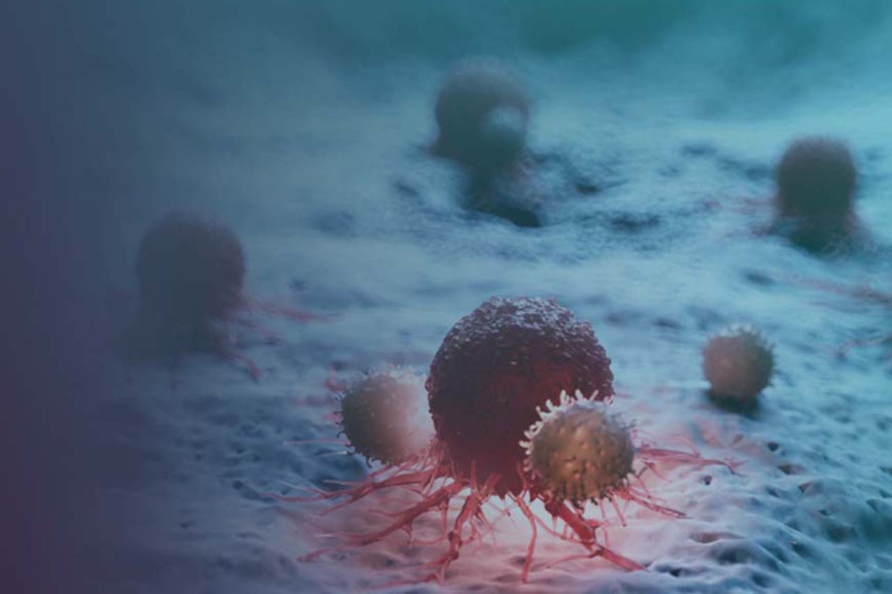
- Immune suppression: Acidic conditions impair T cell, Natural Killer (NK) cell, and dendritic cell activity, undermining the very immune functions that checkpoint inhibitors and other immunotherapies rely on to eliminate tumors.
- Therapy resistance: Acidic pH reduces chemotherapy uptake, especially weak bases, and dampens drug efficacy.
Hypoxia and acidosis are therefore not byproducts but partners in crime that help tumors resist and survive.
The Hidden Force Behind Drug Resistance4
Resistance is often viewed as a genetic issue, driven by mutations in key oncogenes such as EGFR (epidermal growth factor receptor), ALK (anaplastic lymphoma kinase), or KRAS (Kirsten rat sarcoma viral oncogene), that allow tumor cells to bypass or outsmart targeted therapies. But even without such mutations, a hostile TME can undermine treatment. Under acidic conditions, cancer cells reduce drug uptake, reprogram survival pathways, and increase the activity of efflux pumps (specialized transport proteins that actively expel drugs from the cell, preventing them from reaching their targets).
Immunotherapies are equally vulnerable. Checkpoint inhibitors like anti–PD-1 depend on active cytotoxic T cells to recognize and destroy tumor cells. In acidic conditions, however, these T cells lose their effectiveness: interferon-γ secretion decreases, weakening their ability to signal and recruit other immune cells; cytotoxic activity diminishes, reducing direct tumor killing; and populations of myeloid-derived suppressor cells (MDSCs) expand, further silencing the immune response. Together, these changes turn the tumor into an immunologically “cold” environment, one that resists checkpoint blockade and remains effectively invisible to the immune system.
How Hypoxia and Acidosis Enable Immune Escape and Spread3
Low oxygen and low pH reshape both the immune landscape and the physical structure of the tumor. Acidosis impairs antigen presentation, reducing the immune system’s ability to recognize tumor cells; it also drives PD-L1 upregulation, allowing cancer cells to directly suppress T cell activity, and skews dendritic cells toward tolerance rather than immune activation. At the same time, hypoxia remodels the extracellular matrix, loosening structural barriers and clearing pathways that facilitate invasion and metastasis. Together, these forces create a dual advantage for the tumor: they shield it from immune attack while enabling it to spread to new sites.
Strategies to Break the Barrier5,6
To address these challenges, researchers are developing therapies that reprogram the TME:
- Hypoxia-inducible factor (HIF) inhibitors: Drugs that block HIF transcription factors, the “switches” tumors use to adapt to low oxygen by driving angiogenesis, metabolic reprogramming, and survival.
- Monocarboxylate transporter (MCT) blockers: Inhibitors of membrane transport proteins that shuttle lactate and protons out of cancer cells; by blocking them, acid buildup and lactate-driven signaling can be reduced.
- Carbonic anhydrase IX (CAIX) inhibitors: Agents that target CAIX, an enzyme induced under hypoxia that helps tumors regulate pH and survive in acidic environments.
- Systemic buffers: Agents such as sodium bicarbonate that raise blood and tissue pH more generally, but with limited specificity for tumors and potential systemic side effects. By contrast, one approach stands out: L-DOS47, a targeted pharmacological therapy designed specifically to normalize tumor acidity within the tumor microenvironment, offering greater precision and therapeutic potential.
L-DOS47: A Targeted pH Normalizer7
L-DOS47 is a first-in-class antibody-enzyme conjugate (AEC) that delivers a highly localized correction of tumor acidity. It integrates a camelid-derived single-domain antibody (nanobody) targeting CEACAM6 (an adhesion molecule minimally expressed in healthy tissues but overexpressed in many solid tumors, including NSCLC) with a urease enzyme. Once bound to CEACAM6-expressing tumor cells, the urease component becomes catalytically active and converts naturally occurring urea into ammonia and bicarbonate, raising local pH and counteracting acidity.
This localized alkalization leads to:
- Immune reactivation: T cells and NK cells perform better in physiological pH, making L-DOS47 a potential amplifier of PD-1/PD-L1 checkpoint inhibitors.
- Improved drug penetration: Acidity no longer traps weak-base chemotherapy drugs outside tumor cells, improving their uptake.
- Specific targeting: By binding CEACAM6, activity is focused where it’s needed, in tumor tissues, avoiding non-specific systemic alkalization.
Preclinical and clinical evidence is encouraging. In mouse models of pancreatic adenocarcinoma, combining L-DOS47 with the anti-PD-1 antibody pembrolizumab achieved a 70% greater reduction in tumor volume and a 50% greater reduction in tumor weight compared to pembrolizumab alone, within 28 days.(source) Earlier clinical studies in Stage IV NSCLC showed that L-DOS47 combined with pemetrexed and carboplatin improved responses in heavily pre-treated patients, supporting the rationale that normalizing tumor pH can enhance the activity of ionizable, weak-base chemotherapies, such as pemetrexed, and suggesting L-DOS47 is safe and capable of modifying TME acidity in NSCLC patients(source).
Where Does L-DOS47 Fit?
L-DOS47 isn’t meant to replace existing therapies but to unlock their full potential. Its value lies in complementing:
- Checkpoint inhibitors, by transforming resistant, immunologically “cold” tumors into “hot”, immune-inflamed ones.
- Cell therapies (CAR-T, TILs), which currently struggle to persist in acidic environments.
- Next-gen antibodies and ADCs, by improving access and engagement within tumors.
Conclusion
The challenge in NSCLC is not only genetic mutations but also the hostile environment tumors create for themselves. Hypoxia and acidosis shield cancer, suppress immunity, and blunt therapy, and countering these barriers could be the missing key to durable responses.
L-DOS47 represents a groundbreaking, tumor-targeted strategy to normalize pH and restore balance in the TME. By lifting the acidic barrier, it prepares the ground for existing and future treatments to achieve their full potential. As development advances, L-DOS47 could become a pivotal ally in reshaping the NSCLC treatment landscape.
References:
1. Molina JR, Yang P, Cassivi SD, Schild SE, Adjei AA. Non-small cell lung cancer: epidemiology, risk factors, treatment, and survivorship. Mayo Clin Proc. 2008 May;83(5):584-94. doi: 10.4065/83.5.584. PMID: 18452692; PMCID: PMC2718421.
2. Binnewies M, Roberts EW, Kersten K, et al. Understanding the tumor immune microenvironment. Nat Med. 2018;24(5):541–50.
3. Corbet C, Feron O. Tumor acidosis: from the passenger to the driver’s seat. Nat Rev Cancer. 2017;17(10):577–93.
4. Ramalingam SS, Vansteenkiste J, Planchard D, et al. Overall survival with osimertinib in untreated EGFR-mutated NSCLC. N Engl J Med. 2020;382(1):41–50.
5. Wigerup C, Pahlman S, Bexell D. Therapeutic targeting of hypoxia. J Intern Med. 2016;280(6):598–623.
6. Halestrap AP, Wilson MC. The monocarboxylate transporter family—role and regulation. IUBMB Life. 2012;64(2):109–19.
7. Choi Y, Bonfils C, Erlichman C, et al. L-DOS47: a targeted urease immunoconjugate for the modulation of the tumor microenvironment in lung cancer. Cancer Immunol Immunother. 2018;67(11):1879–92.